Retained placenta (also known as retained fetal membranes) is the most common post-partum complication in mares. Typically, expulsion of the placenta occurs shortly after birth and it is considered retained if it is not expelled within 3 hours post-partum. The prevalence of retained placenta varies from 2 to 10% of foalings and can be as high as 30 -54% of uneventful births in Friesian mares. Retention of the placenta in mares should not be overlooked. Appropriate diagnosis and treatment should be quickly applied to prevent secondary, life threatening, complications.
Often retained placenta often occurs following dystocia (difficult birth), prolonged gestation, cesarean section (C-section), fetotomy (obstetrical intervention to remove a dead foal in pieces), hydropsy (excessive amount of the fetal fluids), induced-birth and periparturient hemorrhage of major uterine blood vessels (bleeding). Many factors predispose the mares to retained placenta including: uterine inertia and fatigue, selenium deficiency, calcium/phosphorus imbalances, abnormal hormonal environment, physical (mechanical) intervention during foaling (i.e. dystocia), fescue toxicosis, placentitis, and advanced age. Mares with history of retained placenta have a higher probability of recurrence. Occasionally, retained placenta occurs after an apparently normal foaling for unknown or unidentified reasons.
Overview of the pathophysiology
The mechanisms leading to the retention of the placenta is not completely understood. Following rupture of the umbilical cord at birth, the vessels in the umbilicus and placenta collapse. This along with post-partum uterine contractions moving from the tip of the horns towards the cervix facilitates detachment and eventual infolding of the membranes into the horn so that placenta is normally expelled inside out. The weight of the placenta protruding out from the vulvar lips appears to physically help the expulsion of the placenta.
Retention of the placenta occurs most commonly at the tip of the non-pregnant horn, probably due to the greater connection of the microvilli (microcotyledons), compared to the swollen pregnant horn where the microvilli are more compressed and smaller.
Diagnosis & Treatment
Diagnosis of retained placenta may be obvious in mares having the placenta protruding from the vulva. However, it is more challenging when a small piece of fetal membrane is retained within the uterus. Post-partum placental examination is paramount to verify it is intact, has normal morphology and for early diagnosis of retention of portions of the placenta within the uterus (also called “placental tags”). In those cases where the placenta is not available for inspection, a veterinary practitioner can perform a transrectal ultrasonographic evaluation of the uterus to detect structures in the uterine lumen or attached to the endometrium.
Early diagnosis and treatment of retained placenta or “placental tags” is critical to prevent severe complications such as metritis, septicemia and laminitis, which can start to develop as early as 4 to 8 hours following delivery of the foal. Necrotic fetal membranes within the uterus provide an excellent environment for bacterial growth and endotoxin release. If not treated, retained placenta will progress to septicemia/endotoxemia which can lead to laminitis and death. Metritis (infection of the uterine wall) and endotoxemia (bacterial product) should be suspected in post-partum mares exhibiting fever, anorexia, depression, abnormal mucous membrane color or refill time, tachycardia, muscle twitching and altered manure production.
Local and systemic therapies are used in the treatment of retained placenta. The main goals of the therapy are to: maintain uterine contractility, to decrease uterine inflammation, and to control bacterial growth. Depending upon the clinical status of the mare, she may benefit from referral to a care facility where intensive management can be provided.
Consider Referral if:
- the treatment cannot be applied regularly (e.g. uterine flushing, drug administration)
- signs of sepsis are present (i.e. systemic disease)
- the mare has previous history of laminitis or signs of acute laminitis
- the practitioner lacks experience on handling retained placenta cases
- severe weather may impair daily access to the farm premises
Uterine Lavage
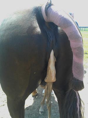
The goal of uterine lavage is three-fold: 1) to physically remove bacteria and cellular debris potentially reducing the risk of secondary metritis, septicemia and laminitis; 2) to stimulate uterine contractility enhancing uterine clearance and perhaps uterine involution; 3) to stimulate neutrophil migration into the uterine lumen to control the infection. Uterine lavage is also particularly useful in cases in which a small piece of fetal membranes is retained. Below (left) is a photo of the retained placenta of a mare after an uneventful parturition and normal gestation. This placenta was retained for 4 days; the mare was treated with antibiotics, ecbolics, anti-inflammatories and daily uterine lavage with a high volume.
Uterine lavage is performed one to three times a day using a large volume (10-20 liters) of Lactated Ringer’s Solution (sterile solution) or tap water (with/without addition of sodium chloride). Uterine lavage is commonly performed using a nasograstic tube. Povidone-iodine may be added to the uterine lavage fluid upon the clinician’s preferences, but caution should be exercised as this antiseptic, when used in high concentrations, may cause endometrial irritation and even necrosis. Mares that underwent a C-section should not have uterine lavage performed due to potential leakage of uterine fluid into the peritoneal cavity and consequently the risk of peritonitis.
The Burns technique is another method that can be used to release retained fetal membranes. Infusing large volume of fluid directly into the allantoic cavity acts to re-expand this space and stretch the attachments of the fetal membranes to the dam’s uterine wall. By expanding the allantoic during the Burns technique, it may remove the tightly held attachments of the placenta with minimal trauma or damage to the mare’s uterus. The difficulty of the technique is obtaining an adequate entrance to the allantoic sac, and this can be difficult if only pieces of the membranes remain within the mare.
Uterotonics
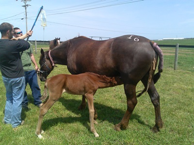
The most common uterotonics (agents used to induce contractions) used to facilitate expulsion of the placenta in broodmares are oxytocin, prostaglandin F 2 alpha (PGF2α) and calcium. Clinically post-partum mares respond to very small doses of oxytocin and signs of discomfort may be observed following administration of even small doses. In the author’s experiences certain breeds such as Morgans, Arabians and Warmbloods and some mares are more likely to show discomfort following oxytocin administration but this can be alleviated, whenever feasible, with hand walking or light sedation (e.g. if the mare cannot be hand walked and she starts kicking that could potentially hurt the foal). Intramuscular injections of oxytocin are preferred as fewer side effects are observed. High doses of oxytocin (>20 units) are not recommended because the ensuing strong uterine contractions are associated with more abdominal discomfort and appear to be less effective in expelling the fetal membranes. In hospitalized mares, oxytocin can be administered as a continued rate infusion, with the expectation of fewer side effects compared to a single bolus injection. A variant of this approach has been extensively applied in the field with good success; this approach consists of slowly administering a solution of oxytocin (0.1 unit/ml in one liter of sterile 0.9% NaCl) over a one hour period. In the photo below (right), a broodmare is receiving intravenous oxytocin treatment for retained placenta while her foal nurses.
Calcium has been recommended for the treatment of retained placenta, especially in Friesian mares (breed known to have the highest incidence of retained placentas). Some veterinarians routinely administer calcium after normal births in draft mares to potentially reduce the risk of placenta retention.
Prostaglandin F 2 alpha and its analog, cloprostenol, are less frequently used to treat retained placenta in mares. Prostaglandin F2 alpha has been demonstrated to have prolonged effects on uterine contractility in non-pregnant cycling mares compared to oxytocin. Similar effects are assumed to occur in mares with retained placenta. Under practical settings, because of its prolonged effect on uterine contractility, PGF2α and its analogs can be used alone or in combination with oxytocin to facilitate the management of retained placenta in situations when multiple oxytocin shots is impractical (e.g. a mare being treated on the farm, evening farm personnel might not be available, etc.).
Physical Manual Removal
 Manual removal of the placenta, either as a prophylactic or therapeutic approach remains controversial. This treatment has the advantage of a quick removal of the fetal membranes. However, the manual removal of the placenta can be associated with several adverse effects such as uterine inversion or prolapse, lacerations, hemorrhage, retention of the micro-villous in the endometrium, delayed uterine involution, increased intrauterine fluid and consequent increasing likelihood of uterine infection. All these effects may induce permanent damage of the endometrium that results in reduced fertility or perhaps chronic infertility. Retention of the micro-villous inside the endometrial crypts can increase the risks of the mare developing metritis. Nonetheless, a recent study reported no significant differences in reproductive performance among mares with retained placenta with or without manual removal of the placenta. Despite the controversy, the author’s current approach is to use very gentle traction (i.e. subjectively only use the tip of the finger without putting much strength to do it) which seems to be somewhat beneficial in some cases without the negative consequences.
Antimicrobials
Mares with retained placentas should be given broad spectrum antimicrobials, with the goal of preventing/reducing bacterial growth, secondary septicemia, and endotoxemia. Several antimicrobial combinations can be used upon specific needs for a mare with retained placenta. However, typically hospitalized mares are administered intravenous antibiotics. On a farm setting, oral antimicrobials can be administered when intravenous drug administration is not feasible.
Once clinical improvement is observed (no evidence of fever, endotoxemia, septicemia or laminitis, and normalization of blood work abnormalities) and the placenta has been expelled the author elects to switch from IV to oral antibiotics. Re-evaluation of the mare is advised before antimicrobial treatment is discontinued to ensure positive response to treatment. Antimicrobials should be administered for 7 to 15 days after the membranes are passed, depending on the clinical status of the mare. In addition, the mare should be re-evaluated within 7 to 21 days post foaling/abortion by transrectal palpation and ultrasound to assure proper uterine involution and to prepare the mare for breeding or to ascertain that the mare is not infected if the owner is not planning to breed her. Broad spectrum, long acting injectable antibiotic can be used when oral antimicrobial administration is not possible. Throughout treatment mares should be monitored for fever, changes in the general demeanor and abnormal vulvar discharge.
Non-steroidal Anti-inflammatories
Non-steroid anti-inflammatory drugs are used in the treatment of mares with retained placenta due to their anti-inflammatory, analgesic and anti-endotoxic effects. Flunixin meglumine is routinely administered to mares with retained placenta as it weakens the deleterious blood flow effects associated with endotoxemia. If the mare is painful due to trauma following dystocia or following surgery, the dose is increased to provide pain relief, although whenever possible the full dose of flunixin meglumine should not be used as it may “mask” fever. Thus, as soon as the signs of inflammation or pain are reduced the dose should be lowered.
Use of phenylbutazone (bute) instead of flunixin meglumine may be beneficial if signs of laminitis are present. Gastrointestinal toxicity (ulceration) is increased when phenylbutazone is administered in combination with flunixin meglumine. Despite potentiated side effects and questionable benefits, this combination is frequently advocated by some practitioners.
Supporting Medications and Procedures
Ideally, mares with retained placentas should have the protruding part of the fetal membranes tight up close to the vulva to avoid the mare stepping on the placenta and cause uterine damage or rip it off and loose the weight effect or avoid the foal sucking on the necrotic fetal membranes which could be a potential source of infection for the neonate. Nonetheless, maiden mares or nervous mares may kick the hanging placenta when it touches their hindquarters thus putting the foal at risk. Exercise is recommended whenever the mare does not have signs of laminitis. A tetanus vaccination should be administered to mares without known vaccination history or mares that are not routinely vaccinated.
 Proton pump inhibitors, such as omeprazole is recommended in critically ill hospitalized mares that may be stressed, have a lack of appetite and prone to gastric irritation and potential ulceration. Such horses are also at risk for gastric irritation and ulceration development when receiving anti-inflammatory therapy, as they may not eat which aggravates the situation.
For mares developing signs of laminitis, oral pentoxifylline is commonly added to the treatment regimen because of its anti-inflammatory action and thought action to improve peripheral blood circulation. It has been postulated that pentoxifylline benefits blood supply to the hoof. Although to date the benefits of pentoxifylline to mares with retained placenta have not been critically evaluated
Hydration status should be closely monitored to prevent complications. Mares showing signs of sepsis will benefit of hospitalization so intravenous fluids can be provided. Intravenous fluid therapy using crystalloid solutions is highly recommended to maintain normal circulating blood volume, to correct dehydration if present, and to help counterbalance the possible effects of endotoxemia.
Pain management is important in these cases, either when the mare presents signs of laminitis or due to painful foaling laceration or necrotic vaginitis following foaling lesions. In hospitalized mares, a constant rate solution of lidocaine (slowly administered intravenously) can be used not only as an analgesic but also as an anti-inflammatory. Painful mares usually have reduced appetite and lay down for prolonged periods. These mares can develop ulcerous and muscular lesions. Such mares may need a digital block with lidocaine (a local anesthetic) in order to get up for feeding and defecation. Mares with retained placenta when kept in stalls must have a deep bed with shavings and grass hay, or sandy floor.
Ice can be applied around the mare’s feet as it has been shown to decrease the severity of laminitis. Hoof cryotherapy can only be effectively achieved by use of commercially available ice boots. If the retained placenta occurred during the winter time where a descent amount of snow is accumulated outside, the mare can be turned out to help decrease the temperature on her feet. If laminitis is suspected, radiographs should be taken to evaluate the severity of the condition.
Secondary complications and prognosis
Future fertility is favorable if the condition is treated in the early stages. Long term prognosis for survival is poor to moderate for mares that develop severe forms of laminitis, endotoxemia and metritis. Laminitis seems to be a more life-threatening condition in draft mares due to their large body weight whereas Quarter Horse mares and related breeds appear to present complications due to proportionally small feet compared to body weight. The prognosis in any mare is determined by the severity of the metritis, septicemia and mainly laminitis. If the mare develops severe metritis the mare may be under risk of death. Again, whenever possible, mares with prolonged retained placenta and presenting laminitis signs (e.g. digital pulse, reluctance to move, prolonged decumbency) should undergo radiograph examination in order to evaluate coffin bone rotation and consequently prognosis.
Take home message
Retained placenta is a potentially life-threating condition to postpartum mares. If not treated early, mares will develop metritis and laminitis. Fortunately, most cases of retained placenta can be easily managed on the farm and require just minimal intervention (e.g. small doses of oxytocin). If the placenta is retained more than 5-6 hours in addition to ecbolics, mares should also receive antibiotics, anti-inflammatories and uterine lavage. Mares with retained placenta, showing signs of sepsis should be referred to care facilities. Specific conditions, where the retained placenta cannot receive appropriate treatment, should also be referred. Mares presenting signs of laminitis should undergo radiograph examination to evaluate phalangeal rotation and prognosis.
_____________________________________________________________________________________
Dr. Igor Canisso is a veterinarian with clinical and research interests in reproductive medicine and obstetrics. He holds a Master’s degree in reproduction. He is a board certified clinician in Animal Reproduction by the American College of Theriogenologists and in Equine Reproduction by the European College of Animal Reproduction. He is interested in all aspects of equine reproduction. Currently, he is a PhD candidate at the Maxwell H. Gluck Equine Research Center, University of Kentucky; soon he will start to serve as an assistant professor of Theriogenology in the Section of Equine Medicine, Department of Veterinary Clinical Medicine at the University of Illinois, Urbana-Champaign. Dr. Canisso has authored two dozen peer review manuscripts and 4 book chapters in veterinary books. Dr. Canisso frequently presents the results of his scientific discoveries and clinical observations in national and international veterinary meetings.

